Want to hear a cool fact about dogs? They have a third eyelid that’s located in the inner corner of the eye under the lower eyelid. Generally, most pet owners don’t notice it unless there is an issue. A condition called cherry eye is a relatively common issue that makes the third eyelid very noticeable. Recognizing cherry eye in your dog and understanding the basic treatment will help keep your dog comfortable if it happens (and prevent you from worrying).
Understanding Cherry Eye in Dogs
A dog’s third eyelid is also called the nictitating membrane. It provides an extra protective layer for the eye and is beneficial when dogs are hunting or in a fight.
At the base of the third eyelid is a gland (nictitans gland) that aids in tear production. The gland can prolapse (pop out) and protrude from the eye if the fibrous attachment (the retinaculum) that anchors it to the lower rim of the eye is weak. This can occur in one or both eyes as a congenital defect or because of injury or trauma to the eye. This condition is called cherry eye. You will notice a pink or red swelling /mass usually at the inside corner of the eye.
When this sensitive tissue is exposed, it’s at risk of drying out and suffering additional trauma. This can result in inflammation, swelling, or infection. Further complications, such as dry eye syndrome (keratoconjunctivitis sicca), can arise if not treated.
This condition is most commonly seen in dogs under the age of two, but can occur in older dogs especially with trauma. Some breeds appear to be more susceptible to cherry eye, including:
- Boston Terriers
- Bulldogs (English or French)
Cocker Spaniels
- Lhasa Apsos
- Pekingese
- Shih Tzus
- Other flat-faced breeds
There is no way to prevent or stop this condition from occurring.
Cherry eyes are uncomfortable and should be seen by your veterinarian. It’s important to have them evaluated by your veterinarian to prevent complications.
What Causes Cherry Eye in Dogs?
The exact cause of a cherry eye is unknown. It may be a result of one or a combination of factors, like:
- Breakdown of supporting structures
- Shallowness of the eye orbit
- Conformation of the eyelids
- Genetics
- Injury or trauma
- Eye infection
Susceptibility to this condition is hereditary. This means that it can be passed from the parents to the offspring. If you’re getting a breed prone to cherry eye, it’s important to ask the breeder if either parent had it.
What Does Cherry Eye Look Like in Dogs?
A cherry eye is located near the inner corner of the eye. It can look like a small pink bulge that may come and go. Generally, owners notice a pinkish-red, fleshy mass about the size of a cherry pit or a bit larger. The longer it is present, the more inflammation occurs, which causes the gland to become irritated and red.
Since it can be itchy, some dogs may paw or rub at their eyes. This can cause the gland to swell more. In some cases, your dog may be unable to completely close their eye due to the size of the gland.
Treatment of Cherry Eye in Dogs
Cherry eyes must be treated as soon as possible. Serious complications can occur if they are left untreated, like:
- Conjunctivitis
- Eye discharge
- Dry eye
Serious complications can occur with dry eye, such as infections and corneal ulcers. This can result in vision loss.
Surgery is the treatment of choice to secure the third eyelid gland behind the third eyelid. Once the gland is secured, inflammation will diminish and prevent another prolapse. Your veterinarian will explain the best surgical technique based on your dog’s condition.
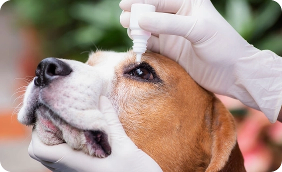
It may be necessary for your veterinarian to prescribe topical antibiotics and/or anti-inflammatories before surgery. This will help treat any infections in the eye and gland as well as relieve some discomfort. In some cases, this can temporarily resolve the prolapse. If the gland is not actively prolapsed, your veterinarian will not be able to perform surgery. Therefore, they will wait until the gland is permanently prolapsed before proceeding.
Following surgery, routine monitoring of your dog’s tear production is important to ensure the gland continues to function properly.

ZPC-02513